HFE (gene)
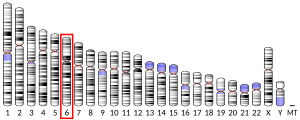
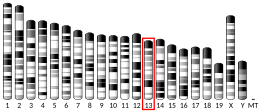
Human homeostatic iron regulator protein, also known as the HFE protein (High FE2+), is a transmembrane protein that in humans is encoded by the HFE gene. The HFE gene is located on short arm of chromosome 6 at location 6p22.2 [5]
Function
[edit]The protein encoded by this gene is an integral membrane protein that is similar to MHC class I-type proteins and associates with beta-2 microglobulin (beta2M). It is thought that this protein functions to regulate circulating iron uptake by regulating the interaction of the transferrin receptor with transferrin.[6]
The HFE gene contains 7 exons spanning 12 kb.[7] The full-length transcript represents 6 exons.[8]
HFE protein is composed of 343 amino acids. There are several components, in sequence: a signal peptide (initial part of the protein), an extracellular transferrin receptor-binding region (α1 and α2), a portion that resembles immunoglobulin molecules (α3), a transmembrane region that anchors the protein in the cell membrane, and a short cytoplasmic tail.[7]
HFE expression is subjected to alternative splicing. The predominant HFE full-length transcript has ~4.2 kb.[9] Alternative HFE splicing variants may serve as iron regulatory mechanisms in specific cells or tissues.[9]
HFE is prominent in small intestinal absorptive cells,[10][11] gastric epithelial cells, tissue macrophages, and blood monocytes and granulocytes,[11][12] and the syncytiotrophoblast, an iron transport tissue in the placenta.[13]
Clinical significance
[edit]The iron storage disorder hereditary hemochromatosis (HHC) is an autosomal recessive genetic disorder that usually results from defects in this gene.
The disease-causing genetic variant most commonly associated with hemochromatosis is p. C282Y.[14] About 1/200 of people of Northern European origin have two copies of this variant; they, particularly males, are at high risk of developing hemochromatosis.[15] This variant may also be one of the factors modifying Wilson's disease phenotype, making the symptoms of the disease appear earlier.[16]
Allele frequencies of HFE C282Y in ethnically diverse western European white populations are 5-14%[17][18] and in North American non-Hispanic whites are 6-7%.[19] C282Y exists as a polymorphism only in Western European white and derivative populations, although C282Y may have arisen independently in non-whites outside Europe.[20]
HFE H63D is cosmopolitan but occurs with greatest frequency in individuals of European descent.[21][22] Allele frequencies of H63D in ethnically diverse western European populations are 10-29%.[23] and in North American non-Hispanic whites are 14-15%.[24]
At least 42 mutations involving HFE introns and exons have been discovered, most of them in persons with hemochromatosis or their family members.[25] Most of these mutations are rare. Many of the mutations cause or probably cause hemochromatosis phenotypes, often in compound heterozygosity with HFE C282Y. Other mutations are either synonymous or their effect on iron phenotypes, if any, has not been demonstrated.[25]
Interactions
[edit]The HFE protein interacts with the transferrin receptor TFRC.[26][27] Its primary mode of action is the regulation of the iron storage hormone hepcidin.[28]
Hfe knockout mice
[edit]It is possible to delete part or all of a gene of interest in mice (or other experimental animals) as a means of studying function of the gene and its protein. Such mice are called “knockouts” with respect to the deleted gene. Hfe is the mouse equivalent of the human hemochromatosis gene HFE. The protein encoded by HFE is Hfe. Mice homozygous (two abnormal gene copies) for a targeted knockout of all six transcribed Hfe exons are designated Hfe−/−.[29] Iron-related traits of Hfe−/− mice, including increased iron absorption and hepatic iron loading, are inherited in an autosomal recessive pattern. Thus, the Hfe−/− mouse model simulates important genetic and physiological abnormalities of HFE hemochromatosis.[29] Other knockout mice were created to delete the second and third HFE exons (corresponding to α1 and α2 domains of Hfe). Mice homozygous for this deletion also had increased duodenal iron absorption, elevated plasma iron and transferrin saturation levels, and iron overload, mainly in hepatocytes.[30] Mice have also been created that are homozygous for a missense mutation in Hfe (C282Y). These mice correspond to humans with hemochromatosis who are homozygous for HFE C282Y. These mice develop iron loading that is less severe than that of Hfe−/− mice.[31]
HFE mutations and iron overload in other animals
[edit]The black rhinoceros (Diceros bicornis) can develop iron overload. To determine whether the HFE gene of black rhinoceroses has undergone mutation as an adaptive mechanism to improve iron absorption from iron-poor diets, Beutler et al. sequenced the entire HFE coding region of four species of rhinoceros (two browsing and two grazing species). Although HFE was well conserved across the species, numerous nucleotide differences were found between rhinoceros and human or mouse, some of which changed deduced amino acids. Only one allele, p.S88T in the black rhinoceros, was a candidate that might adversely affect HFE function. p.S88T occurs in a highly conserved region involved in the interaction of HFE and TfR1.[32]
See also
[edit]Notes
[edit]
The 2015 version of this article was updated by an external expert under a dual publication model. The corresponding academic peer reviewed article was published in Gene and can be cited as: James C Barton, Corwin Q Edwards, Ronald T Acton (9 October 2015). "HFE gene: Structure, function, mutations, and associated iron abnormalities". Gene. Gene Wiki Review Series. 574 (2): 179–192. doi:10.1016/J.GENE.2015.10.009. ISSN 0378-1119. PMC 6660136. PMID 26456104. Wikidata Q30380172. |
References
[edit]- ^ a b c GRCh38: Ensembl release 89: ENSG00000010704 – Ensembl, May 2017
- ^ a b c GRCm38: Ensembl release 89: ENSMUSG00000006611 – Ensembl, May 2017
- ^ "Human PubMed Reference:". National Center for Biotechnology Information, U.S. National Library of Medicine.
- ^ "Mouse PubMed Reference:". National Center for Biotechnology Information, U.S. National Library of Medicine.
- ^ "HGNC: HFE". Retrieved 30 August 2019.
- ^ "NCBI Gene: HFE homeostatic iron regulator". National Center for Biotechnology Information. Retrieved 30 November 2020.
 This article incorporates text from this source, which is in the public domain.
- ^ a b Feder JN, Gnirke A, Thomas W, Tsuchihashi Z, Ruddy DA, Basava A, et al. (August 1996). "A novel MHC class I-like gene is mutated in patients with hereditary haemochromatosis". Nature Genetics. 13 (4): 399–408. doi:10.1038/ng0896-399. PMID 8696333. S2CID 26239768.
- ^ Dorak MT (March 2008). "HFE (hemochromatosis)". Atlas of Genetics and Cytogenetics in Oncology and Haematology. Archived from the original on 29 September 2017. Retrieved 17 June 2020.
- ^ a b Martins R, Silva B, Proença D, Faustino P (March 2011). "Differential HFE gene expression is regulated by alternative splicing in human tissues". PLOS ONE. 6 (3): e17542. Bibcode:2011PLoSO...617542M. doi:10.1371/journal.pone.0017542. PMC 3048171. PMID 21407826.
- ^ Waheed A, Parkkila S, Saarnio J, Fleming RE, Zhou XY, Tomatsu S, et al. (February 1999). "Association of HFE protein with transferrin receptor in crypt enterocytes of human duodenum". Proceedings of the National Academy of Sciences of the United States of America. 96 (4): 1579–1584. Bibcode:1999PNAS...96.1579W. doi:10.1073/pnas.96.4.1579. PMC 15523. PMID 9990067.
- ^ a b Griffiths WJ, Kelly AL, Smith SJ, Cox TM (September 2000). "Localization of iron transport and regulatory proteins in human cells". QJM. 93 (9): 575–587. doi:10.1093/qjmed/93.9.575. PMID 10984552.
- ^ Parkkila S, Parkkila AK, Waheed A, Britton RS, Zhou XY, Fleming RE, et al. (April 2000). "Cell surface expression of HFE protein in epithelial cells, macrophages, and monocytes". Haematologica. 85 (4): 340–345. PMID 10756356.
- ^ Parkkila S, Waheed A, Britton RS, Bacon BR, Zhou XY, Tomatsu S, et al. (November 1997). "Association of the transferrin receptor in human placenta with HFE, the protein defective in hereditary hemochromatosis". Proceedings of the National Academy of Sciences of the United States of America. 94 (24): 13198–13202. Bibcode:1997PNAS...9413198P. doi:10.1073/pnas.94.24.13198. PMC 24286. PMID 9371823.
- ^ Hsu CC, Senussi NH, Fertrin KY, Kowdley KV (August 2022). "Iron overload disorders". Hepatology Communications. 6 (8): 1842–1854. doi:10.1002/hep4.2012. PMC 9315134. PMID 35699322.
- ^ "Hemochromatosis". Archived from the original on 18 March 2007. Retrieved 20 August 2009.
- ^ Gromadzka G, Wierzbicka DW, Przybyłkowski A, Litwin T (September 2022). "Effect of homeostatic iron regulator protein gene mutation on Wilson's disease clinical manifestation: original data and literature review". The International Journal of Neuroscience. 132 (9): 894–900. doi:10.1080/00207454.2020.1849190. PMID 33175593. S2CID 226310435.
- ^ Porto G, de Sousa M (2000). Barton JC, Edwards CQ (eds.). Variation of hemochromatosis prevalence and genotype in national groups. In: Hemochromatosis: Genetics, pathophysiology, diagnosis and treatment: Cambridge University Press. pp. 51–62. ISBN 978-0521593809.
- ^ Ryan E, O'keane C, Crowe J (December 1998). "Hemochromatosis in Ireland and HFE". Blood Cells, Molecules & Diseases. 24 (4): 428–432. doi:10.1006/bcmd.1998.0211. PMID 9851896.
- ^ Acton RT, Barton JC, Snively BM, McLaren CE, Adams PC, Harris EL, et al. (2006). "Geographic and racial/ethnic differences in HFE mutation frequencies in the Hemochromatosis and Iron Overload Screening (HEIRS) Study". Ethnicity & Disease. 16 (4): 815–821. PMID 17061732.
- ^ Rochette J, Pointon JJ, Fisher CA, Perera G, Arambepola M, Arichchi DS, et al. (April 1999). "Multicentric origin of hemochromatosis gene (HFE) mutations". American Journal of Human Genetics. 64 (4): 1056–1062. doi:10.1086/302318. PMC 1377829. PMID 10090890.
- ^ Merryweather-Clarke AT, Pointon JJ, Shearman JD, Robson KJ (April 1997). "Global prevalence of putative haemochromatosis mutations". Journal of Medical Genetics. 34 (4): 275–278. doi:10.1136/jmg.34.4.275. PMC 1050911. PMID 9138148.
- ^ Merryweather-Clarke AT, Pointon JJ, Jouanolle AM, Rochette J, Robson KJ (2000). "Geography of HFE C282Y and H63D mutations". Genetic Testing. 4 (2): 183–198. doi:10.1089/10906570050114902. PMID 10953959.
- ^ Fairbanks VF (2000). Barton JC, Edwards CQ (eds.). Hemochromatosis: population genetics. In: Hemochromatosis: Genetics, pathophysiology, diagnosis and treatment. Cambridge University Press. pp. 42–50. ISBN 978-0521593809.
- ^ Acton RT, Barton JC, Snively BM, McLaren CE, Adams PC, Harris EL, et al. (2000). "Geographic and racial/ethnic differences in HFE mutation frequencies in the Hemochromatosis and Iron Overload Screening (HEIRS) Study". Ethnicity & Disease. 16 (4): 815–821. PMID 17061732.
- ^ a b Edwards CQ, Barton JC (2014). Greer JP, Arber DA, Glader B, List AF, Means Jr RT, Paraskevas F, Rodgers GM (eds.). Hemochromatosis. In: Wintrobe's Clinical Hematology. Wolters Kluwer/Lippincott Williams & Wilkins. pp. 662–681. ISBN 9781451172683.
- ^ Feder JN, Penny DM, Irrinki A, Lee VK, Lebrón JA, Watson N, et al. (February 1998). "The hemochromatosis gene product complexes with the transferrin receptor and lowers its affinity for ligand binding". Proceedings of the National Academy of Sciences of the United States of America. 95 (4): 1472–1477. Bibcode:1998PNAS...95.1472F. doi:10.1073/pnas.95.4.1472. PMC 19050. PMID 9465039.
- ^ West AP, Bennett MJ, Sellers VM, Andrews NC, Enns CA, Bjorkman PJ (December 2000). "Comparison of the interactions of transferrin receptor and transferrin receptor 2 with transferrin and the hereditary hemochromatosis protein HFE". The Journal of Biological Chemistry. 275 (49): 38135–38138. doi:10.1074/jbc.C000664200. PMID 11027676.
- ^ Nemeth E, Ganz T (2006). "Regulation of iron metabolism by hepcidin". Annual Review of Nutrition. 26: 323–342. doi:10.1146/annurev.nutr.26.061505.111303. PMID 16848710.
- ^ a b Zhou XY, Tomatsu S, Fleming RE, Parkkila S, Waheed A, Jiang J, et al. (March 1998). "HFE gene knockout produces mouse model of hereditary hemochromatosis". Proceedings of the National Academy of Sciences of the United States of America. 95 (5): 2492–2497. Bibcode:1998PNAS...95.2492Z. doi:10.1073/pnas.95.5.2492. PMC 19387. PMID 9482913.
- ^ Bahram S, Gilfillan S, Kühn LC, Moret R, Schulze JB, Lebeau A, et al. (November 1999). "Experimental hemochromatosis due to MHC class I HFE deficiency: immune status and iron metabolism". Proceedings of the National Academy of Sciences of the United States of America. 96 (23): 13312–13317. Bibcode:1999PNAS...9613312B. doi:10.1073/pnas.96.23.13312. PMC 23944. PMID 10557317.
- ^ Levy JE, Montross LK, Cohen DE, Fleming MD, Andrews NC (July 1999). "The C282Y mutation causing hereditary hemochromatosis does not produce a null allele". Blood. 94 (1): 9–11. doi:10.1182/blood.V94.1.9.413a43_9_11. PMID 10381492. S2CID 12722648.
- ^ Beutler E, West C, Speir JA, Wilson IA, Worley M (2001). "The hHFE gene of browsing and grazing rhinoceroses: a possible site of adaptation to a low-iron diet". Blood Cells, Molecules & Diseases. 27 (1): 342–350. doi:10.1006/bcmd.2001.0386. PMID 11358396.
Further reading
[edit]- Dorak MT, Burnett AK, Worwood M (March 2002). "Hemochromatosis gene in leukemia and lymphoma". Leukemia & Lymphoma. 43 (3): 467–477. doi:10.1080/10428190290011930. PMID 12002748. S2CID 26047470.
- Beutler E (May 2003). "The HFE Cys282Tyr mutation as a necessary but not sufficient cause of clinical hereditary hemochromatosis". Blood. 101 (9): 3347–3350. doi:10.1182/blood-2002-06-1747. PMID 12707220.
- Ombiga J, Adams LA, Tang K, Trinder D, Olynyk JK (November 2005). "Screening for HFE and iron overload". Seminars in Liver Disease. 25 (4): 402–410. doi:10.1055/s-2005-923312. PMID 16315134. S2CID 25457332.
- Distante S (2006). "Genetic predisposition to iron overload: prevalence and phenotypic expression of hemochromatosis-associated HFE-C282Y gene mutation". Scandinavian Journal of Clinical and Laboratory Investigation. 66 (2): 83–100. doi:10.1080/00365510500495616. PMID 16537242. S2CID 23644937.
- Zamboni P, Gemmati D (July 2007). "Clinical implications of gene polymorphisms in venous leg ulcer: a model in tissue injury and reparative process". Thrombosis and Haemostasis. 98 (1): 131–137. doi:10.1160/th06-11-0625. PMID 17598005. S2CID 33271776.
External links
[edit]- HFE+protein,+human at the U.S. National Library of Medicine Medical Subject Headings (MeSH)
- Overview of all the structural information available in the PDB for UniProt: Q30201 (Hereditary hemochromatosis protein) at the PDBe-KB.


